facs buffer flow cytometry
Perform red blood cell lysis per lab protocol either ACT ACK or LSM. Place samples in 12 x 75 mm Falcon tubes and analyze by flow cytometry as soon as possible within 1 hour.

Flow Cytometry Protocol For Staining Membrane Associated Proteins In Suspended Cells R D Systems
Stable and minimal spillover.
. Add 01-10 μgml of the primary labeled antibody. The exosome pellet was subsequently dissolved in 500 μL buffer. Absence of these ions reduces cation-dependent cell to cell adhesion and prevents clumping.
Also compare to BioLegend buffers to the equivalent BD products. 2 Add 100 μl of 200 μgml DNase-free RNaseA and incubate at 37C for 30 minutes. BSA and FBS or any other serum for that matter will accomplish pretty much the same thing when staining cells for flow.
FACS Buffer we use has 1 BSA and 01 Sodium Azide. Our high-performing flow cytometers equip you for a broad of range of clinical and research applications including measurement of CD4 counts in HIV patients residual white blood cells. Incubate for at least 20-30 min at room temperature of 4C.
Prepare the following buffer in which to suspend cellular samples prior to cell sorting. One important way to minimize non-specific staining is by the use of a so-called blocking reagent. People use protein containing buffers for flow cytometry is to prevent cells from sticking to the side of plastic tubes or other culture labware as well as preventing cell.
Ad NovaFluor dyes designed for spectral flow cytometers. Prepare single-cell suspensions from either lymphoid tissue bone marrow peripheral blood or cell cultures using standard protocols. Our Flow Cytometry Staining Buffer is designed for use in immunofluorescent staining protocols of cells in suspension.
Wash the cells 3 times by centrifugation at 400 g for 5 min and resuspend them in ice. Easy-to-add into multi-color experiments. BioLegend develops and manufactures world-class.
Staining buffer is the buffer used. View available buffers for various flow cytometry applications. Sample preparation reagents for flow cytometry include cell surface staining intracellular and transcription factor staining buffer sets cell lysis assays blocking reagents and magnetic cell.
Harvest wash the cells single cell suspension and adjust cell number to a concentration of 1-5106 cellsml in ice cold FACS. Flow Cytometry Staining Buffer FACS Buffer This basic FACS Buffer is a buffered saline solution that can be used for immunofluorescence staining protocols antibody and cell dilution. 858 784-8396 flowstaffscrippsedu Sort Buffer The proper design of sort buffer for.
Make sure products are not expired. Flow Cytometry Scripps Research 10550 North Torrey Pines Road IMM-20 La Jolla CA 92037 tel. Flow Cytometry Direct immunofluorescence staining.
Flow cytometry FACS staining protocol Cell surface staining 1. This Flow Cytometry Staining. Resuspend cells with 052 mL FACS buffer.
Alternatively samples can be. Use this buffer also for all washes until directed to use Sorting Buffer Adjust. Here are 5 ingredients to consider for your FACS buffer.
Stable and minimal spillover. Ensure that antibodies are stored as per the instructions of manufacturer. Flow Cytometry FACS Blocking.
RBCs are disproportionately permeable. Originally developed in the late 1960s flow cytometry is a popular analytical cell-biology technique that utilizes light to count and profile cells in a heterogenous fluid mixture. Basic Sorting Buffer 1 x Phosphate Buffered Saline PBS or Hanks Balanced Salt Solution HBSS.
Ad NovaFluor dyes designed for spectral flow cytometers. A blocking reagent contains a high concentration of. Preparation of magnetic beads and antibody immobilization on the beads.
3 Add 100 μl of 1 mgml propidium iodide light sensitive and incubate at room temperature for 5-10. Keep track of antibody stocks. Harvest wash the cells and adjust cell suspension to a concentration of 1-5 x 10 6 cellsmL in.
We use this buffer for surface staining as well as for intracellular staining. Sheath Fluid and FACS Buffer Dulbuccos wo Ca2 Mg2 20X for a longer shelf life than 1X or even 10X. General procedure for flow cytometry using a conjugated primary antibody.
If titrating antibodies and storing aliquots of the. This incubation must be done in the dark. Our FACS buffer is based on PBS and contains 2 FCS 005 Sodium Azide.
20X PBS Stock Solution. 1- Use CaMg2 free PBS. Dilutions if necessary should be made in FACS buffer.
Dissolve in THIS ORDER theyll dissolve faster in 1 liter of. The analyses were performed. Incubate for at least 30 min at room temperature or 4C in the dark.
Re-suspend in FACS staining buffer. Easy-to-add into multi-color experiments. PBMC Preparation for Flow Cytometry These protocols are meant to be modified with your experiment specifics in mind.
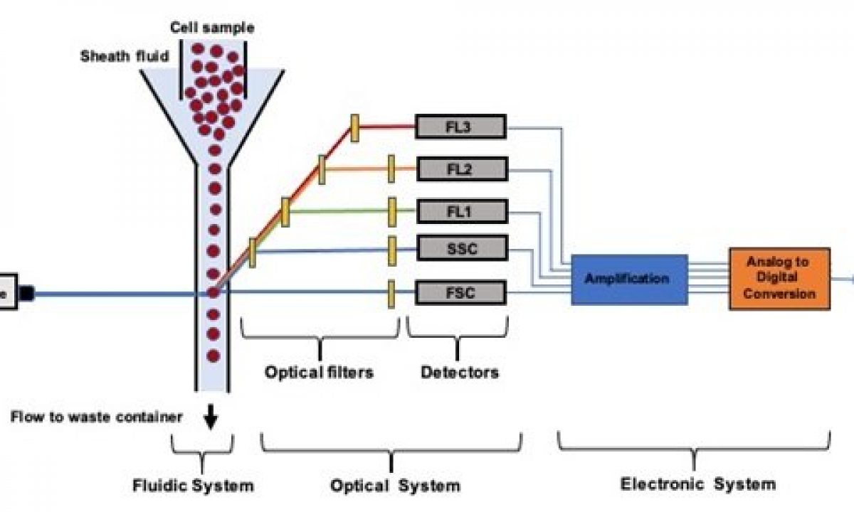
Microfluidic Flow Cytometry Principles And Commercial Review Ufluidix
Facs Buffer Composition

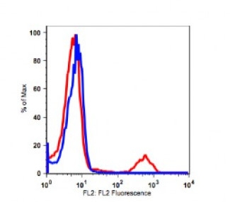
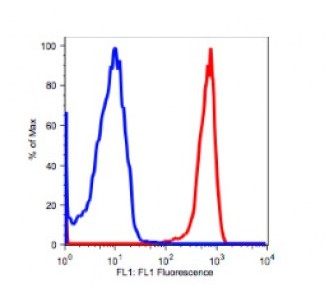
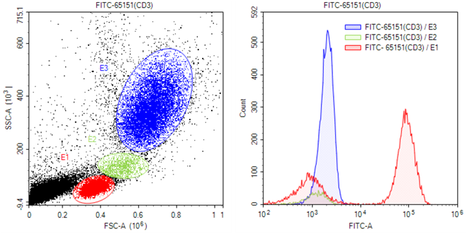
We Offer Anti Mouse Cd3 Antibody Biot Conjugated Keep As Concentrated Solution Store At 4 C And Protected From Prolonged Exposure To Life Science Index Biot

Fundamentals Of Flow Cytometry Aat Bioquest

What Is Flow Cytometry Technology Networks

Flow Cytometry Protocols
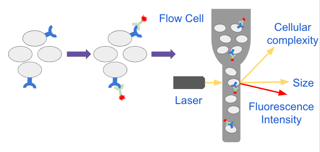
Analyzing Single Cells With Flow Cytometry

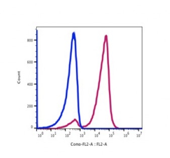
Ep2 Polyclonal Antibody Calculated Mw 40 Kda Observed Mw 40 Kda Source Rabbit Isotype Igg For More Information Click On Life Science Observation Index
Facs Buffer Composition
Facs Buffer Composition
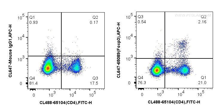
Popular Antibodies For Flow Cytometry Proteintech Group

Pin On Myalgic Encephalomyelitis Chronic Fatigue Syndrome

Flow Cytometry And Cell Sorting By Facs In The Flow Cell 1 The Download Scientific Diagram

Flow Cytometry Control And Standardization Beads
Flow Cytometry And Cell Sorting By Facs In The Flow Cell 1 The Download Scientific Diagram
Flow Cytometry Perm Buffer 10x Pf00011 C Proteintech
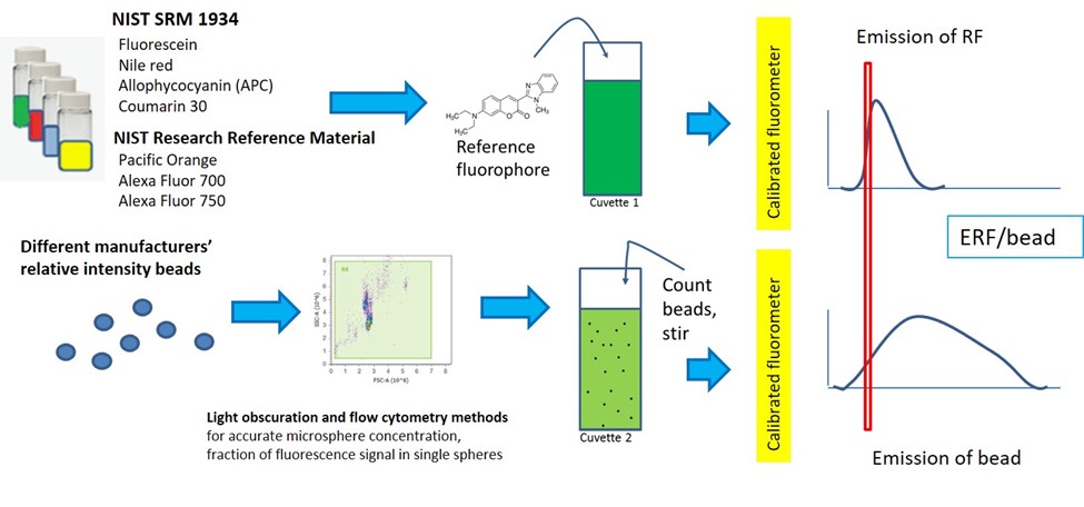
Quantitative Flow Cytometry Measurements Nist
Facs Buffer Composition

Key Steps In Flow Cytometry Protocols